Anatomy and Function of the Pons
Parts of the Pons:
The pons is an approximately 2cm long “knob-like” process located deep in the middle of the brain.
One can visualize the pons as a box…
- Top – Midbrain
- Bottom – Medulla
- Front – Pontine Cistern and the Clivus (the slanted bony bottom part of the skull)
- Back – 4th ventricle
- Sides – Cerebellar Peduncles
Contents
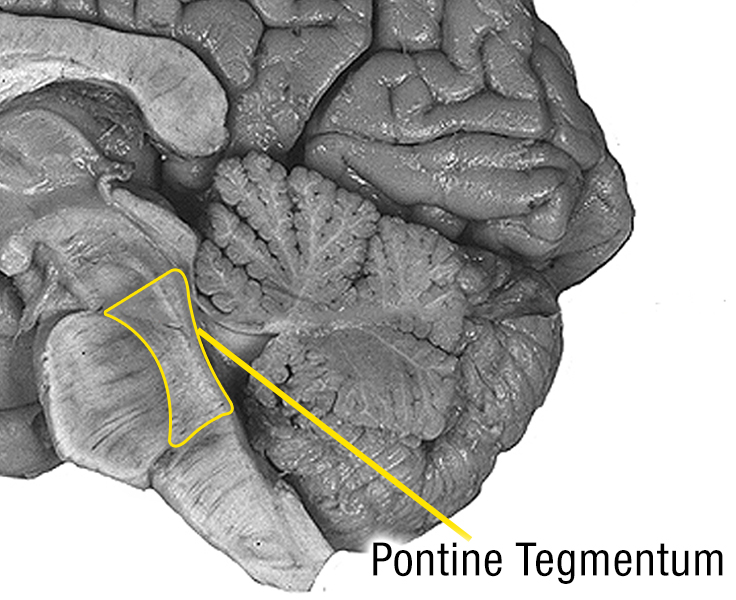
The pons itself is divided into a back (medically called dorsal) part and a front (medically called ventral) part. The back part is often called the Pontine Tegmentum whereas the front part is called the Ventral or Basal Pons. There are 4 paired cranial nerves (one on the left and one on the right) that start in the pons.
Pontine Tegmentum:
The pontine tegmentum is against the 4th ventricle and contains an origin of 4 cranial nerves (5th, 6th, 7th and 8th nerve), the reticular activating system, ascending/descending nerve tracts, as well as other nuclei.
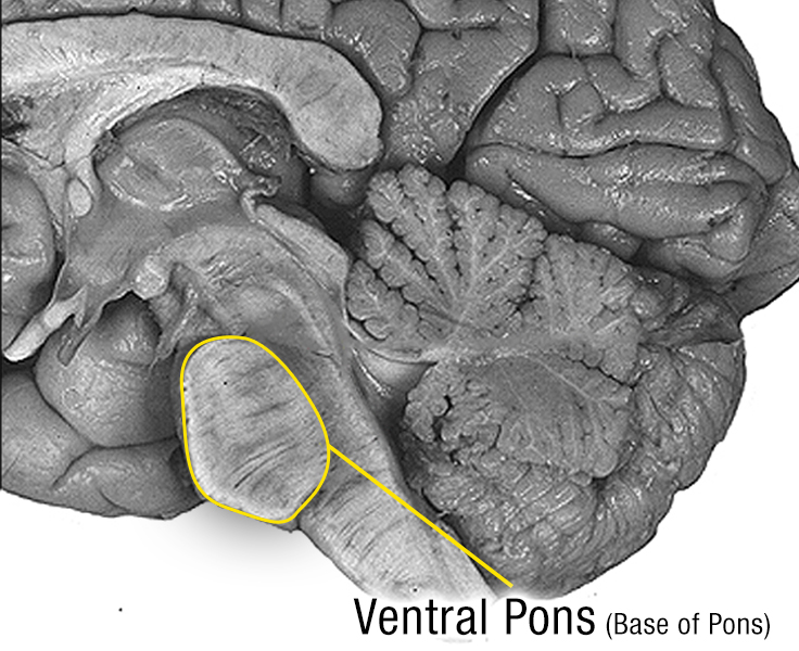
Ventral Pons:
The ventral pons consists of nerve fiber tracts going up and down as well as across the area. It acts as a massive relay station connecting the cerebral cortex to the cerebellum on the other side through a structure called the cerebellar peduncles.
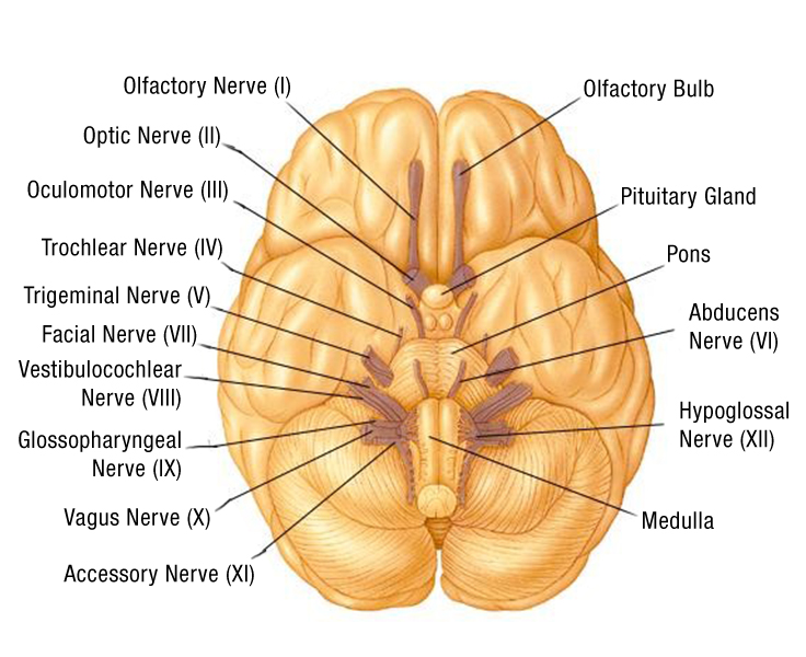
Cranial Nerves:
The cranial nerves are nerves that control the special senses and the movement/sensation of the head and neck.
Four sets of cranial nerves come from the pons.
5th – Trigeminal nerve
6th – Abducens nerve
7th – Facial nerve
8th – Vestibulocochlear nerve

The Brainstem
of which the pons is the upper part, has three main functions
Allowing nerve bundles to travel back and forth from the cerebrum to the cerebellum as well as nerve bundles to and from the body. Essentially all information from the brain to the body or vice versa must go through someplace in the brainstem.
The cranial nerves that originate in the pons are the:
- 5th nerve – Trigeminal – This nerve has a sensory and motor component.
- The sensory part is responsible for the feeling of the face.
- The motor part innervates the muscles of the mandible responsible for biting, chewing and swallowing.
- 6th nerve – Abducens – This is a motor nerve that allows to eye to look to the side. Dysfunction of the nerve will cause the eye to drift in towards the nose potentially eventually becoming paralyzed in this position.
- 7th nerve – Facial – This is a motor nerve that affects the muscles of facial expression such as smiling, showing one’s teeth, raising the eyebrows, closing the eyes tightly or puffing out the cheeks.
- More information about facial paralysis
- 8th nerve – Vestibulocochlear – This is a sensory nerve with two parts.
- The cochlear portion of this nerve allows for hearing transmitted sound from the ear to the brain. Problems with this part of the nerve can cause hearing loss.
- The vestibular portion transmits information from the inner ear about one’s position in space which allows for balance and coordination. Dysfunction causes vertigo, motion sickness, nystagmus and a loss of equilibrium.
- Consciousness – The reticular formation is the dorsal part of the midbrain and brainstem (in the tegmentum). It is involved in the sleep/wake cycle affecting fatigue, alertness and motivation. Some theorize that the reticular formation has a role in dreaming.
- Respiration – There are two centers in the pons that affect respiration.
- The apneustic center in the lower pons seems to stimulate and prolong inspiration thereby controlling the intensity of breathing.
- The pneumotaxic center located in the upper pons inhibits inspiration. It decreases the depth and frequency of breaths.
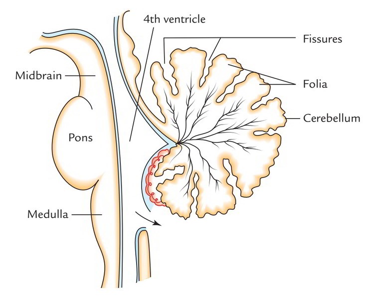
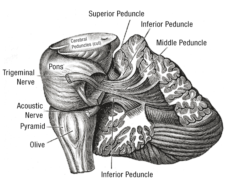
Cerebellar Peduncles:
The cerebellar peduncles (also called brachium pontis) are the nerve superhighway from the pons to the cerebellum.
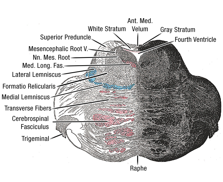
Microscopic Cross Section of the Pons:
The area above the blue is the tegmentum.
The area below the blue is the ventral pons.
The crossing fibers in the vental pons continue to the sides to form the cerebellar peduncles.
Blood Supply:
The blood to the pons is supplied by the pontine arteries. These are small arteries that branch off the basilar artery.
Used with permission from Just One More Day
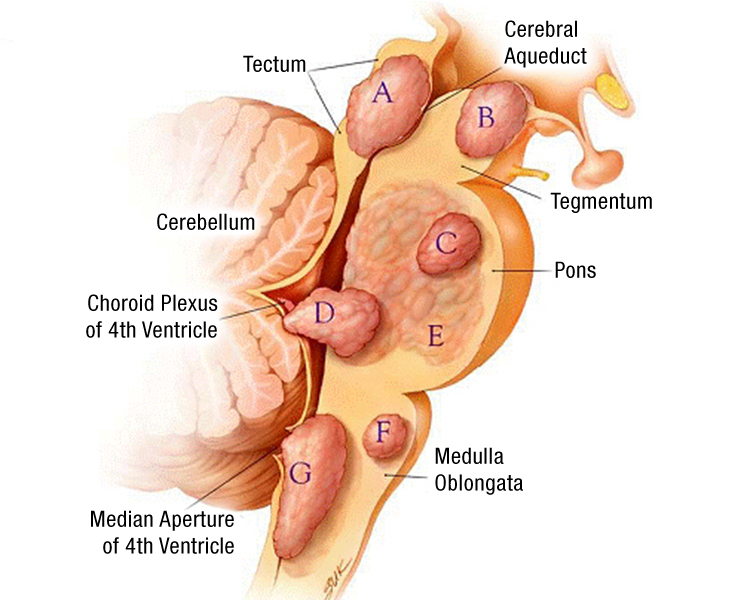
Different Brainstem Glioma Locations
A- Tectal Glioma
B- Focal midbrain tumor
C- Focal intrinsic pontine glioma
D- Doral/exophytic glioma
E- Diffuse Intrinsic Pontine Glioma
F- Focal medullary glioma
G- Cervicomedullary glioma